Introduction
The assessment of cartilage health is of paramount significance in radiology, as cartilage plays an important role in joint features and mobility. an expansion of imaging modalities has been advanced to assess cartilage fitness, providing particular insights into its shape and integrity. In this article, we delve into the numerous imaging strategies utilised in radiology to assess cartilage health, highlighting their strengths, limitations, and packages.
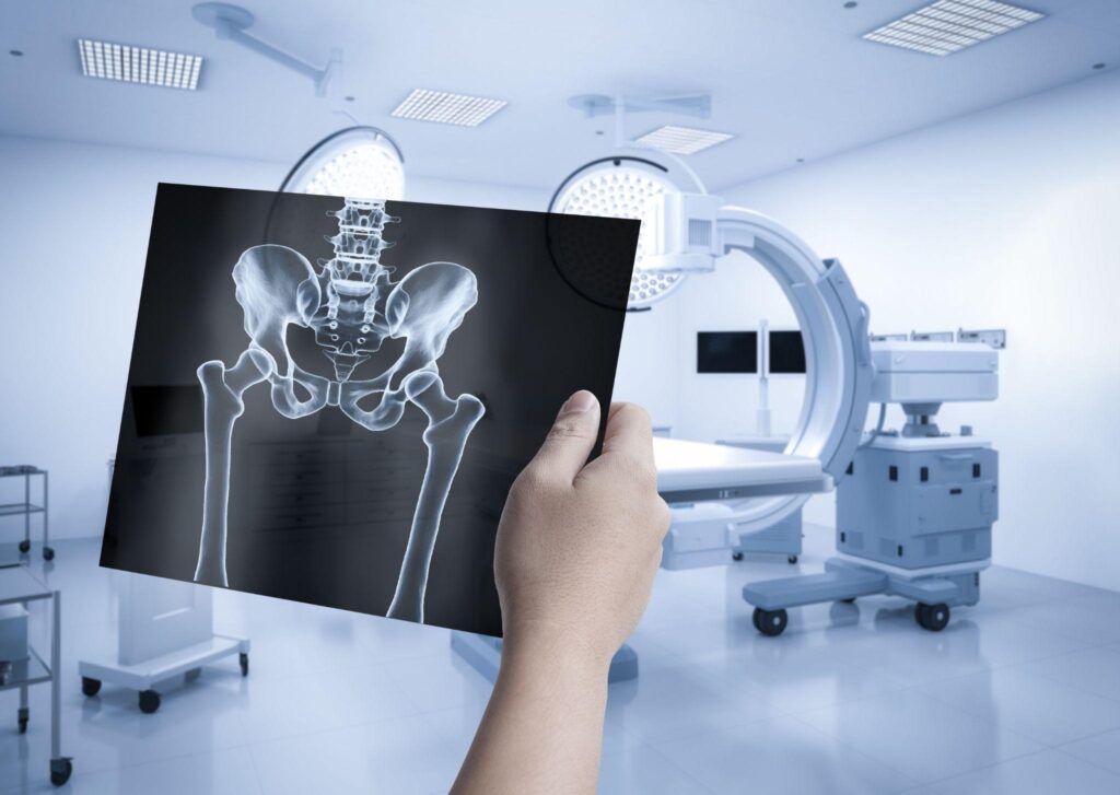
X-ray Imaging
X-ray Imaging: Revealing Cartilage Health Indirectly
X-ray imaging, a cornerstone of medical imaging, employs electromagnetic radiation to generate images of bones and underlying structures. While it might not directly visualise cartilage, this time-tested technique plays a crucial role in assessing cartilage health through indirect indicators. By capturing changes in joint spaces, bone structure, and osteophyte formation, X-ray imaging provides valuable insights into cartilage degeneration, making it an essential tool in diagnosing musculoskeletal conditions, particularly advanced osteoarthritis.
Principle of X-ray Imaging:
X-ray imaging functions by emitting controlled doses of X-rays, a type of electromagnetic radiation, through the body. These rays are absorbed differently by various tissues, with bones appearing as white areas on X-ray images due to their higher density, while softer tissues like cartilage appear darker. Cartilage itself doesn’t absorb X-rays well, which is why it’s challenging to visualise directly using this method. However, the changes that occur in bone and joint structures due to cartilage deterioration can be observed through X-ray imaging.
Indirect Indicators of Cartilage Degeneration:
While cartilage remains invisible on X-ray images, the effects of its degeneration are readily apparent. As cartilage deteriorates, the space between bones in a joint narrows due to the loss of cushioning material. This narrowing of the joint area is a key signal of cartilage damage and is a primary characteristic observed through X-ray imaging. Furthermore, because the cartilage wears away, the frame responds by forming bony growths known as osteophytes, which are seen on X-rays as irregular protrusions at the rims of the bones.
Diagnostic importance:
X-ray imaging’s capability to not directly display cartilage health is mainly precious in diagnosing superior osteoarthritis. In cases where significant cartilage loss has occurred, the joint space narrows significantly, exposing bones to increased friction and potential damage. The presence of osteophytes further confirms the degenerative changes in the joint. This information aids physicians in making accurate diagnoses, formulating treatment plans, and providing necessary interventions to manage pain and improve patient quality of life.
Limitations and Considerations:
While X-ray imaging provides valuable insights, it has limitations. It’s not suitable for detecting early cartilage degeneration or assessing soft tissues directly. Moreover, X-rays involve exposure to ionising radiation, which poses potential risks, especially with frequent use. In situations where more detailed cartilage assessment is required, other imaging modalities like MRI or ultrasound may be more appropriate.
X-ray imaging, despite its inability to directly visualise cartilage, remains an indispensable tool for evaluating cartilage health. By capturing changes in joint spaces, bone structures, and osteophyte formation resulting from cartilage degeneration, X-ray imaging aids in diagnosing advanced osteoarthritis and other musculoskeletal conditions. Its role in identifying indirect indicators of cartilage health underscores its significance in providing valuable information for healthcare professionals and contributing to effective patient care.
Magnetic Resonance Imaging (MRI)
Magnetic Resonance Imaging (MRI): Revolutionising Cartilage Assessment
Magnetic Resonance Imaging (MRI) has emerged as a cornerstone in medical imaging, offering unparalleled insights into soft tissues. When it comes to evaluating cartilage health, MRI stands out as a versatile and comprehensive modality. Its ability to provide detailed images and specific cartilage-related sequences makes it an exceptional tool for diagnosing and monitoring various cartilage conditions.
MRI’s Capabilities in Cartilage Assessment:
At the heart of MRI lies its capability to capture high-decision pictures of tender tissues, making it especially well-applicable for assessing cartilage health. In contrast to X-ray imaging or CT scans, MRI no longer depends on ionising radiation, making it more secure for repeated use. Specialised sequences like T2-weighted imaging highlight the water content of cartilage, aiding in the identification of early cartilage degeneration. Additionally, T1rho mapping reveals the biochemical composition of cartilage, assisting in detecting subtle changes before they manifest visibly.
Quantitative Analysis and Cartilage Thickness:
One of MRI’s standout features is its capability to perform quantitative analysis of cartilage health. By utilising specific techniques, MRI can not only visualise cartilage but also measure its thickness accurately. This record is priceless for diagnosing situations where cartilage thinning occurs, which includes osteoarthritis. The capability to music modifications in cartilage thickness over time allows scientific professionals to screen ailment development and tailor remedy techniques accordingly.
Multiplanar Imaging for Comprehensive Views:
MRI’s multiplanar capabilities empower radiologists to obtain comprehensive views of cartilage defects and lesions. This means that not only can cartilage be assessed from various angles, but the surrounding structures can also be thoroughly evaluated. This level of detail aids in making accurate diagnoses, understanding the extent of cartilage damage, and planning for surgical interventions when necessary.
Advancements in Cartilage Imaging:
MRI’s contributions to cartilage assessment continue to expand with ongoing advancements. Techniques like dynamic contrast-enhanced MRI provide insights into cartilage perfusion and metabolism, enhancing our understanding of cartilage health beyond its structure alone. Additionally, 3D imaging techniques offer improved visualisation and quantification of cartilage morphology, further refining diagnostic accuracy.
Challenges and Considerations:
While MRI is an effective tool, it’s no longer without boundaries. get right of entry to MRI machines can be confined, and the procedure may be time-consuming. Patients with certain medical devices or conditions won’t be eligible for MRI. in addition, the price of MRI scans and the need for specialised education in interpretation are critical elements to bear in mind.
Magnetic Resonance Imaging (MRI) has revolutionised the manner we determine cartilage health. Its detailed imaging capabilities, coupled with specialised sequences like T2-weighted and T1rho mapping, offer insights into early cartilage degeneration, quantify thickness, and evaluate the biochemical composition. MRI’s multiplanar capabilities enable comprehensive views of cartilage defects, enhancing diagnostic accuracy and treatment planning. As technology continues to advance, MRI remains a crucial tool in the field of musculoskeletal imaging, contributing to improved patient care and outcomes.
Computed Tomography (CT) Imaging
Computed Tomography (CT) Imaging: Peering into Cross-Sectional Detail
Computed Tomography (CT) Imaging has brought intricate cross-sectional visualisation to the realm of medical imaging, offering detailed insights into the human body’s internal structures. While it primarily excels in visualising bones, CT can be harnessed as part of a multi-modal approach for assessing cartilage health. This article delves into the unique role CT plays in the evaluation of cartilage and its associated considerations.
CT Imaging Mechanics:
CT imaging makes use of X-rays to generate a chain of go-sectional pics, which are then reconstructed into detailed 3-D images of the frame’s interior. The method presents radiologists with a comprehensive view of bones, organs, and tissues, facilitating unique diagnoses and remedy plans.
Cartilage Assessment in CT:
Although CT imaging is renowned for its bone imaging capabilities, it can play a valuable role in assessing cartilage health, especially when combined with other modalities. CT arthrography is a technique that enhances cartilage visualisation by injecting a contrast agent directly into the joint space. This contrast agent helps outline the cartilage surfaces, aiding radiologists in identifying abnormalities.
Limitations in Cartilage Evaluation:
While CT imaging can provide a wealth of information about bones and some insight into cartilage, it has limitations when it comes to cartilage assessment. Cartilage is not well-contrasted in standard CT scans due to its low density and poor X-ray attenuation. As a result, CT is less sensitive to subtle changes in cartilage structure and composition compared to techniques like MRI.
Radiation Exposure and Monitoring:
One significant consideration in CT imaging is the exposure to ionising radiation, which carries potential risks, particularly with repetitive use. This radiation exposure makes CT less suitable for ongoing monitoring of cartilage health, especially in conditions that require frequent imaging.
Role in analysis and remedy planning:
CT imaging, whilst hired along with other modalities, can provide treasured facts in diagnosing joint situations that involve both bone and cartilage. It can help identify the extent of joint degeneration, assess bone quality, and provide a broader context for cartilage-related issues.
Computed Tomography (CT) Imaging offers a powerful cross-sectional view of the body’s structures, particularly bones, that can be complemented with specialised techniques for cartilage assessment. While not the primary choice for directly visualising cartilage, CT arthrography and its ability to enhance cartilage visualisation add a valuable dimension to the evaluation of joint health. However, the limitations in sensitivity to subtle cartilage changes and the potential for radiation exposure should be carefully considered when deciding on the most appropriate imaging modality for each clinical scenario.
Ultrasound Imaging
Ultrasound Imaging: Unveiling Cartilage Health in Real Time
Ultrasound imaging has emerged as a dynamic and handy imaging modality that employs sound waves to create actual-time snapshots of inner systems. At the same time as it is extensively diagnosed for obstetric and cardiac imaging, its talents make it bigger for musculoskeletal assessment, together with the assessment of cartilage health. This article delves into the prowess of ultrasound in this regard, highlighting its strengths and limitations.
Precept of Ultrasound Imaging:
Ultrasound imaging operates on the precept of emitting high-frequency sound waves into the frame. These waves jump off inner structures, inclusive of cartilage, and are then processed to create pictures in actual-time. This real-time capability provides physicians with immediate insights, making ultrasound a valuable tool for various medical scenarios.
Cartilage Assessment through Ultrasound:
Ultrasound’s versatility extends to evaluating cartilage health, providing a non-invasive approach to visualising this vital joint component. It has the ability to assess cartilage thickness, and surface irregularities, and detect cartilage lesions. In instances of joint trauma or degenerative situations, ultrasound can provide a real-time view of cartilage harm.
Advantages of Ultrasound:
Ultrasound’s advantages are manifold. It’s cost-effective, widely accessible, and doesn’t expose patients to ionising radiation, making it suitable for repeated assessments. Its real-time imaging capabilities are especially valuable when monitoring joint movements and functionality. Additionally, it can be used in dynamic assessments, such as evaluating joint mobility and cartilage response to loading.
Limitations and Operator Dependency:
However, ultrasound’s accuracy is significantly dependent on the operator’s skill and experience. The quality of images obtained can vary, impacting diagnostic confidence. Additionally, ultrasound is better suited for assessing superficial joints due to its limited penetration capabilities. Deeper joints might present challenges in obtaining clear images of cartilage structures.
Role in Sports Medicine and Point-of-Care Settings:
Ultrasound’s real-time imaging and dynamic capabilities have made it a go-to tool in sports medicine. It enables direct visualisation of cartilage health during joint movement, aiding in understanding the impact of various activities on cartilage. Moreover, its portability makes it suitable for point-of-care evaluations, where quick assessments are essential.
Ultrasound imaging stands as a dynamic and accessible modality for evaluating cartilage health. Its real-time capabilities, cost-effectiveness, and ability to assess cartilage thickness, and surface irregularities, and detect lesions make it a valuable tool in musculoskeletal imaging. While its accuracy can vary based on operator skill and its limited penetration may pose challenges, ultrasound remains a crucial asset in sports medicine, point-of-care settings, and instances where immediate insights into cartilage health are paramount.
Nuclear Medicine Techniques: Unveiling Cartilage Metabolism and Viability
In the realm of medical imaging, nuclear medicine techniques have introduced a unique dimension by harnessing the power of radioactive substances to visualise metabolic activity within the body. While not as widely recognized as traditional imaging methods, nuclear medicine techniques offer valuable insights into cartilage health by focusing on its metabolism, perfusion, and even viability. This article delves into the significance of these techniques, shedding light on their capabilities and applications in the realm of cartilage assessment.
Principles of Nuclear Medicine:
Nuclear medicine techniques involve the administration of trace amounts of radioactive substances, known as radiotracers, into the body. These radiotracers emit gamma rays, which are detected by specialised cameras during imaging. The emitted gamma rays provide information about the metabolic processes occurring within tissues, thereby offering functional insights that can’t be gleaned from structural imaging alone.
Functional Insights into Cartilage:
Two main nuclear medicine techniques – Single-photon Emission Computed Tomography (SPECT) and Positron Emission Tomography (PET) – provide invaluable functional insights into cartilage metabolism and perfusion. These techniques allow physicians to gauge the activity levels of tissues, including cartilage, shedding light on the biochemical processes that underlie its health.
Sodium MRI and NaF-PET: Exploring Cartilage Viability:
In addition to metabolism and perfusion assessments, more experimental techniques like Sodium Magnetic Resonance Imaging (MRI) and Positron Emission Tomography with Sodium Fluoride (NaF-PET) have emerged to evaluate cartilage viability and detect early changes in conditions like osteoarthritis. These techniques provide insights into the cellular health of cartilage, allowing for a deeper understanding of its state.
Strengths and Considerations:
Nuclear medicine techniques offer unique strengths in assessing cartilage health. By focusing on metabolic activity, they can identify functional abnormalities before structural changes become apparent. However, these techniques have limitations, including the need for specialised equipment, the potential for radiation exposure, and the challenges in interpreting functional data.
Integration for Comprehensive Assessment:
Each imaging modality, from X-ray and MRI to ultrasound and nuclear medicine techniques, has its strengths and weaknesses. The choice of modality depends on factors such as the suspected condition, the joint being assessed, and the desired level of detail. Employing a combination of techniques can offer a comprehensive understanding of cartilage health, enabling accurate diagnosis and tailored treatment planning.
Nuclear medicine techniques bring a unique perspective to cartilage assessment by revealing its metabolic activity, perfusion, and viability. While more experimental and less commonly used, these techniques provide functional insights that complement traditional structural imaging methods. By integrating these techniques into the diagnostic approach, radiologists can enhance their ability to accurately diagnose cartilage-related conditions and develop targeted treatment strategies for optimal patient outcomes.
Conclusion
The evaluation of cartilage health is a crucial aspect of radiology, enabling early detection and management of cartilage-related conditions. Each imaging modality brings its own set of advantages and limitations to the table, allowing radiologists to tailor their approach based on the clinical context and the specific goals of assessment. X-ray imaging provides a broad view of joint changes, while MRI offers detailed information about cartilage morphology and biochemical composition. CT arthrography enhances cartilage visualisation, ultrasound provides real-time assessment, and nuclear medicine techniques offer insights into cartilage metabolism. As technology continues to advance, the integration of these modalities promises more accurate and comprehensive evaluations of cartilage health, ultimately improving patient care and outcomes.